How to position and take TPLO Radiographs
Two views are required for measurements of a TPLO surgery
Positioning of the patient
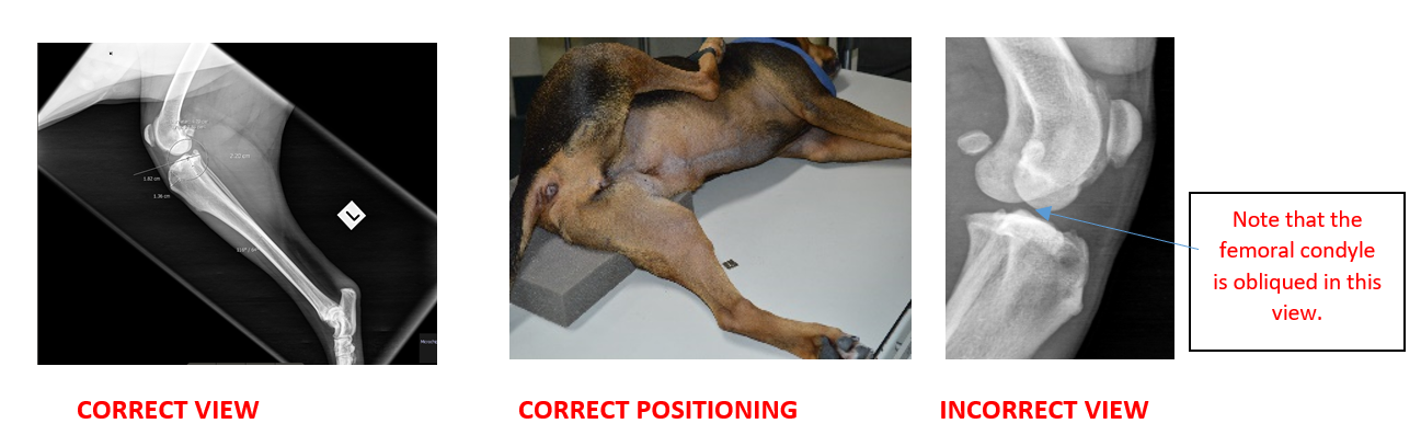
1st View
Lateral Radiograph
The patient should be lying in lateral recumbancy with the leg of interest closest to the table/plate.
Place a foam pad under the pelvis to elevate it.This will result in a better lateral position of the distal femur. The lateral and medial femoral condyles should be superimposed. Collimate to include stifle and hock.
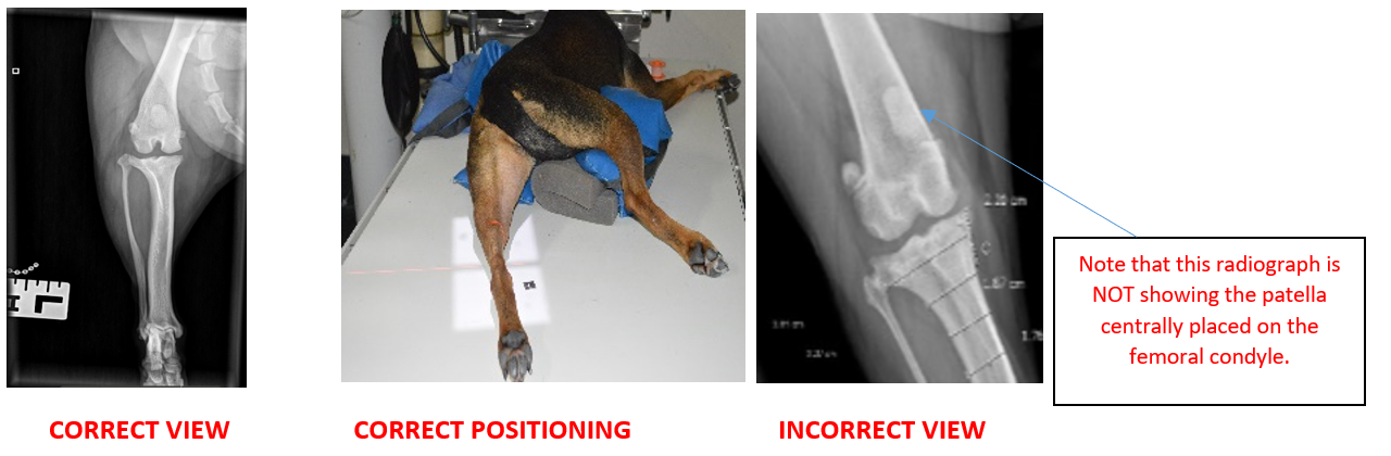
2nd View
Craniocaudal Radiograph
Patient needs to be lying in sternal recumbency with the leg of interest extending caudally.
Elevate the opposite leg with foam and/or sandbags to remove from view. The pelvis should be slightly rolled towards the affected limb. Make sure that the leg of interest is not rotated.
Ensure that L/R markers are included on all radiographs.